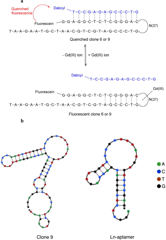
Fig. 1.
a Design adopted to measure the binding between the selected clones (clone 6 and 9) and Gd(III) ion. Clone 6 and 9 is tagged with a fluorophore (fluorescein) and hybridized with a 13-base long QS bearing a quencher (dabcyl). In the absence of Gd(III), the clone-QS hybrid is quenched. Adding Gd(III) leads to displacement of QS and the formation of clone-Gd(III) complex, which results in fluorescence growth. b Structures of clone 9 and the truncated Ln-aptamer as predicted using NUPACK [23]. The ‘loop’ region of clone 9 is conserved in Ln-aptamer, and the latter is subsequently adapted into a sensor for detecting the presence of free Gd(III) ion following the strategy depicted in a

