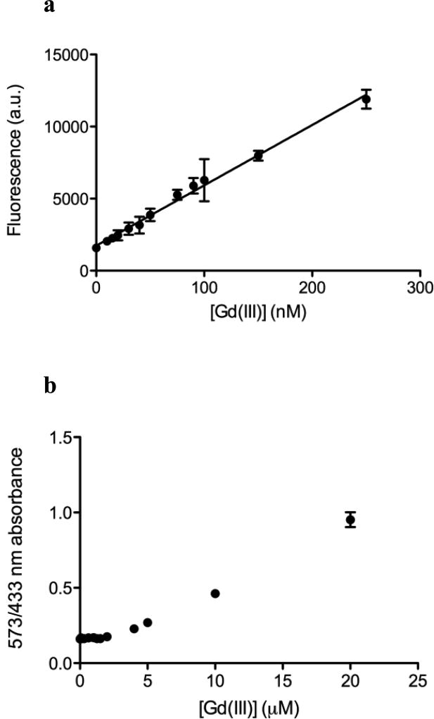
Fig. 4.
a Fluorescence change of Ln-aptamer with increasing concentration of aqueous Gd(III) ion. The response is linear from 0 to 250 nM of Gd(III). R2 = 0.9935, P < 0.0001 (linear regression on Prism 5.0, Graphpad Software). The limit of detection (S/N = ~3) is at ~80 nM of Gd(III). All data are plotted as average raw fluorescence reading in arbitrary units (a.u.) ± standard deviation, n = 6. b Detection of aqueous Gd(III) ion using xylenol orange. The limit of detection is found to be ~10 – 15 µM (S/N = ~3) based on the change in ratio of absorbance at 573 and 433 nm

