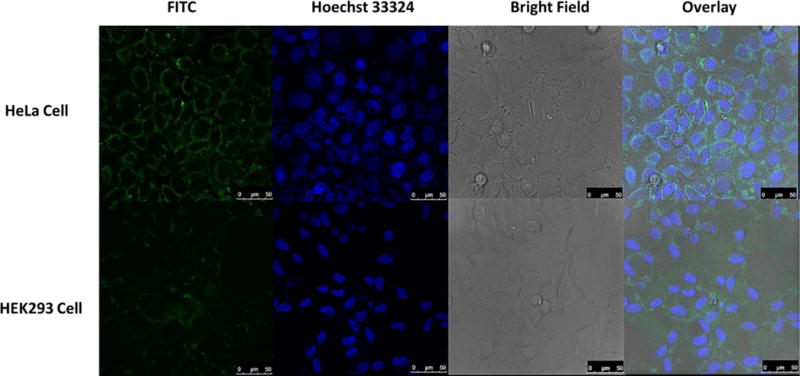
Figure 4.

Confocal fluorescence images of living HeLa cells and HEK293 cells incubated with AS1411-2 conjugate. Each series can be sorted as FITC fluorescence, nucleus of cells dyed in blue by Hoechst 33324, optical images of cells in bright field and the overlay of the previous channels, respectively. All scale bars are 50 μm.
