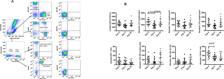
Fig 1.
(A) Gating scheme for phenotype analyses of the various cell markers in the peripheral blood from a representative animal. (B) Relocation-dependent differences in lymphocytes in rhesus monkeys. The lymphocytes were first gated based on forward scatter (FCS) versus side scatter (SSC), and then CD3+ T cells, CD3-CD16+ natural killer (NK) cells, CD3+CD16+ NKT cells, and CD20+ B cells were positively identified. Further analyses of CD3+ T cells showed CD4+ T cells, CD8+ T cells, and CD4+CD8+ double-positive T cells. The specificity of staining for the various markers was ascertained according to the isotype control antibody staining used for each pair of combination markers, as shown.
Aliquots of EDTA whole blood were stained with fluorescence-labeled antibodies to the CD3+, CD4+, CD8+, CD20+, and CD16+ lymphocytes and analyzed for T-cell subpopulations in rhesus monkeys. Values on the Y-axis are absolute lymphocytes cells. P values were considered statistically significant at p<0.05.

