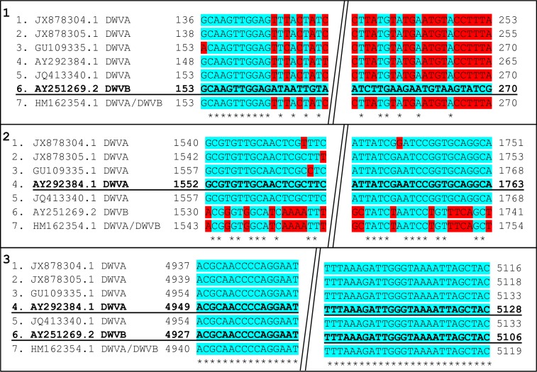
Fig 1. Location of quantification viral primers.
1) DWV-B primer location, 2) DWV-A primer location, 3) Pan-DWV primer location, shown on all seven viral sequences used. Bold and Underlined sequences identify which sequence was used for primer design, numbers correspond to location on genome sequences (Forward primers on the left, Reverse on the right), blue highlighted (*) areas indicate consensus and red areas indicate mismatch locations, diagonal double line break indicates removed sequence section between primer locations.

