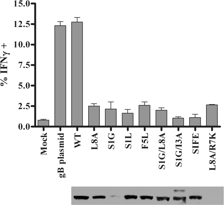
Fig 1. Mutant gB proteins and recognition by gB-CD8s.
Untreated B6WT3 cells (mock) or cells transfected with plasmids to express WT gB or gB498-505 epitope mutants were incubated for eighteen hours. The labeling of the mutations is as depicted in Table 1; WT is a plasmid which went through the mutagenesis procedure without changes. Cells were harvested for expression analysis by immunoblotting using a monoclonal gB-specific antibody (lower panel) or, in parallel, transfected cells were combined with 5x10⁴ gB-CD8s from an endogenously expanded clone and stimulated for 5 h in the presence of Brefeldin A. gB-CD8s were surface stained for CD45 and CD8, permeabilized, and stained for intracellular IFNγ. The graph depicts one of two representative experiments, with the mean percent of IFNγ+ cells (n = 2/group) and standard error of the mean (SEM) for each stimulation.

