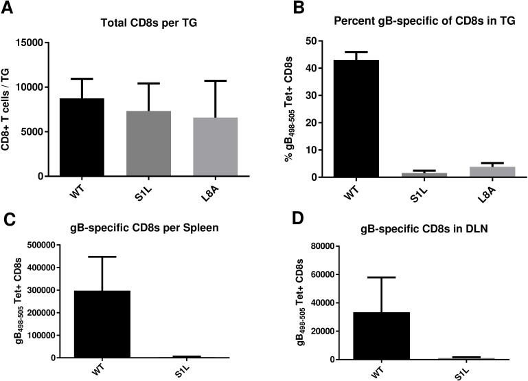
Fig 4. Acute CD8+ T cell infiltrates in the ganglia of mice after corneal infection with WT HSV-1 or recombinant HSV-1 containing gB498-505 mutations.
Corneas of mice were infected with 1x10⁵ PFU/eye of HSV-1 WT, S1L, or L8A. At 8 dpi (peak CD8+ T cell infiltrate), the TG, spleen, or DLN were dissociated into single cell suspensions and surface stained with antibodies to CD45, CD3, CD8 and with MHC-I gB498-505 tetramer as detailed in Methods. Cells were analyzed by flow cytometry, and the data are presented as the mean +/- SEM (n = 5 mice, 10 TGs) of (A) absolute number of CD3+CD8+ T cells per TG, (B) the percent of gB498-505 tetramer positive CD8+ T cells in each TG, or (C, D) the total number of gB498-505 tetramer specific cells per spleen and local DLN. The experiment shown is representative of three additional experiments, all producing similar results. The absolute numbers of CD8+ T cells induced in the TG with each virus were not statistically different as shown by a one-way ANOVA followed by Tukey’s posttest (p = 0.58).

