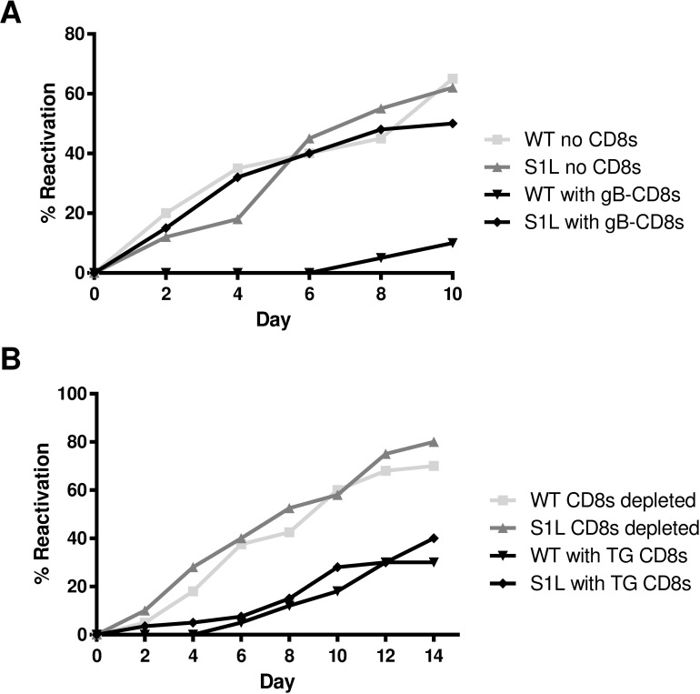
Fig 5. Ex vivo ganglionic reactivation of WT and S1L HSV-1.
Corneas of B6 mice were infected with 1x10⁵ PFU of WT or S1L HSV-1. At 34 dpi latently infected TGs were dispersed with collagenase. (A) The TG cells were depleted at >95% of endogenous CD8+ T cells and distributed to wells of a 48-well tissue culture plate (0.2 TG equivalent/well) and cultured in culture medium containing IL-2 and with or without 2 x 10⁴ gB-CD8s added per well. Culture fluid samples were removed and replaced with fresh media every two days. The presence of infectious virus in culture fluid (indicating HSV-1 reactivation) was then determined by plaque assay. (B) TG cells were mock depleted or depleted of 95% of endogenous CD8+ T cells by treatment with anti-CD8α antibody and complement, then distributed to wells of a tissue culture plate and cultured as described in A above. (A & B) Data plotted as total percentage of wells that reactivated (showing infectious virus in culture supernatant) at the indicated time of culture. n = 10 TG per condition. Data for each experiment are representative of one of two repeats, but experiment-to-experiment variability in reactivation rates are routinely observed.

