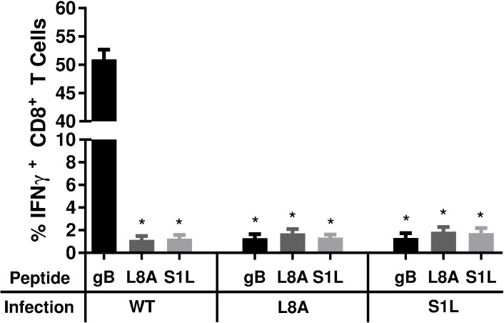
Fig 6. Stimulation of acute TG-resident CD8+ T cell populations with WT, SIL, or L8A gB peptides.
B6 mice received corneal infections with HSV-1 expressing WT, S1L, or L8A gB. TG were obtained at 8 dpi, dispersed into single cell suspensions, and the endogenous CD8+ T cells were stimulated for 6 hours with B6WT3 fibroblasts pulsed individually with WT, S1L, or L8A gB498-505 peptides, in the presence of brefeldin A. Cells were surface stained for CD45 and CD8, followed by an intracellular stain for IFNγ. The data are represented as the mean percentage of CD8+ T cells that produced IFNγ +/- SEM (n = 5 mice per group). * represents significance of p<0.0001 for each group compared to gB peptide stimulation of wild-type infected control (first column) using one-way ANOVA with Dunnett’s multiple comparisons posttest.

