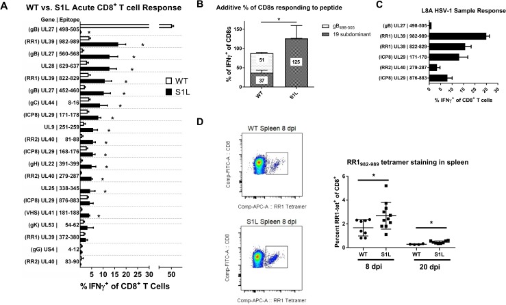
Fig 9. Subdominant HSV-1 epitopes expand to accommodate the loss of the immunodominant gB498-505 epitope during acute infiltration into the TG.
B6 mice received corneal infections with (A, B, D) HSV-1 WT, S1L, or (C) L8A. TG were excised at 8 dpi, dispersed into single cell suspensions, stimulated for 6 hrs in the presence of Brefeldin A with B6WT3 cells pulsed with peptides corresponding to known HSV-specific CD8+ T cell epitopes, stained for surface CD45, CD8, and intracellular IFNγ. (A) The graph shows the percent of the total CD8+ T cell population staining for intracellular IFNγ by flow cytometry. The bars represent the mean ± SEM frequency of CD8+ T cells producing IFNγ in response to each epitope. (B) Total fraction of gB498-505 or non-gB-CD8s responding to peptide stimulations as seen in (A). N = 3–8 TG equivalents per peptide. (C) A fraction of the peptide library was analyzed as in (A), but in mice infected with HSV-1 L8A. (D) Spleens were also excised at 8 and 20 dpi and examined for RR1982-989 tetramer positive cells. Shown is example of tetramer staining, and a graph depicting the total fraction of splenic CD8+ T cells that stained positive for tetramer. A t-test was performed for each matched pair of responding CD8+ T cells, and * denotes p<0.05.

