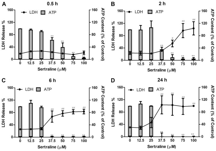
FIG. 1.
Effects of sertraline on cellular ATP level (bars) and cell death (lines) in rat primary hepatocytes. Rat primary hepatocytes were treated with DMSO as vehicle control and sertraline at the concentrations of 0–100μM for 0.5, 2, 6, and 24 h. Cellular ATP content and LDH release were measured as described in the Materials and Methods section. #p < 0.01 and ##p < 0.001 represent LDH release is significantly different from the control for each time point; **p < 0.001 represents ATP depletion is significantly different from the control for each time point. Data are represented as mean ± SD from at least three independent experiments.

