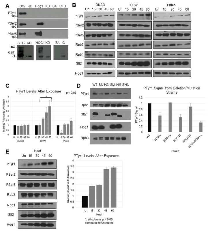
Figure 4. Slt2 phosphorylates Tyr1 in vitro and in vivo.
(A) In vitro kinase assay using 3HA-tagged Slt2 and Hog1 extracted and IPed from activated cells. Following incubation with GST-CTD, proteins were resolved by SDS-PAGE and blots probed with the indicated antibodies. Lanes are Slt2-HA (SLT2) and kinase-dead (KD), Hog1-HA (HOG1) and kinase dead (KD), beads and antibody control (BA) and GST-CTD alone (CTD). (B) Western blot of Tyr1P levels after stress induction. DMSO, calcofluor white (CFW) and phleomycin (Phl) are shown. Rpb1, Slt2, Hog1, and Rpb3 were also probed using their respective antibodies. Time points are untreated (Un), 15, 30, 45 and 60 minutes. (C) Quantification of (B). Signals were normalized individually to Rpb3 levels, then collectively to uninduced control. (D) Western blot and quantification of Tyr1P levels in isogenic deletion strains. Protein extracts from strains with either SLT2/HOG1 deletions (SΔ/HΔ; double, SHΔ) or kinase-dead mutations (SM/HM) were blotted using 3D12 and normalized to WT. (E) Western blot of Tyr1P levels after heat stress (37°C); signals normalized as in (B). All results shown are representative of three independent experiments; data are represented as mean +/− SE of three independent experiments.

