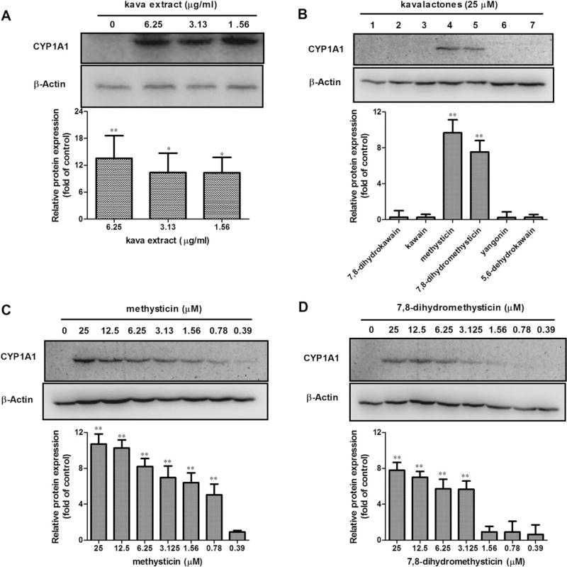
FIG. 3.
Protein expression induction of CYP1A1 by kava extract and kavalactones in Hepa1c1c7. (A) Induction of CYP1A1 by kava extract (0–6.25 µg/ml). (B) Induction of CYP1A1 by six kavalactones (25µM). 1–7 represent DMSO control, 7,8-dihydrokawain, kawain, methysticin, 7,8-dihydromethysticin, yangonin, and 5,6-dehydrokawain, respectively. (C) Induction of CYP1A1 protein expression by methysticin. (D) Induction of CYP1A1 by 7,8-dihydromethysticin. Hepa1c1c7 cells were exposed kava or kavalactones for 24 h. Cell lysates (40 µg) were analyzed by western blot for levels of CYP1A1. All the data are generated from at least three independent experiments. The ratios of CYP1A1 protein against β-actin were calculated; values shown are mean ± SD. *p < 0.05; **p < 0.01 versus control.

