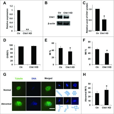
Figure 2.

Chk1 depletion induced oocytes to be arrested at the MI stage and impaired the spindle organization and chromosome alignment. (A) Expression of Chk1 mRNA in the siRNA-injected oocytes. Porcine COCs were microinjected with the control and Chk1-specific siRNA, respectively. After injection, porcine COCs were incubated with dbcAMP for 20 h and followed by qRT-PCR. (B) Expression of Chk1 protein in the siRNA-injected oocytes. Porcine COCs were injected with siRNA and incubated with dbcAMP for 20 h, followed by Western blotting. The molecular mass of Chk1 is 54 kDa and that of β-actin is 42 kDa. (C) Relative levels of Chk1 protein between control and Chk1 knockdown groups. (D) Percentage of GVBD oocytes in the control group and Chk1 siRNA microinjected group. (E) Percentage of MI oocytes in the control group and Chk1 siRNA microinjected group. (F) Percentage of MII oocytes in the control group and Chk1 siRNA microinjected group. (G) Chk1 depletion triggered abnormal chromosomes arrangement; (Blue ovals or circulars showed chromosomes arrangement. The macroaxis of ovals referred to the co-orientation of chromosomes. Circulars also showed that the co-orientation was perpendicular to viewing plane. Bar = 10 μm. (H) Percentage of abnormal MI oocytes in the control group and Chk1 siRNA microinjected group. All graphs show as mean ± SE. *p<0.05. (KD, knockdown).
