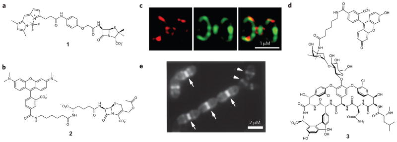
Figure 3. Antibiotic-inspired probes and images obtained with these compounds.
(a,b) Structures of PBP imaging probes. Probe 1 (a) labels all PBPs that are present in bacteria, whereas molecule 2 (b) selectively targets only a subset of PBPs in B. subtilis and S. pneumoniae. (c) 3D-SIM images of PBPs in S. pneumoniae after dual labeling with 2 (red) and 1 (green). (d) Vancomycin-fluorescein (3) enables visualization of nascent PG synthesis. (e) Nascent PG in S. pneumoniae cells is labeled with molecule 3. Arrows indicate heavily stained division septum. Arrowheads indicate lightly stained equators of the daughter cells that will become division sites. Figures reproduced with permission from: c, ref. 45; e, ref. 49.

