
Figure 4.
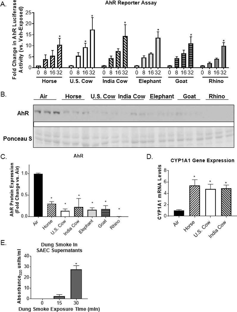
Dung smoke contains AhR ligands and activates the AhR.
(A) A cell line containing an AhR luciferase reporter was treated with the indicated concentrations of dung biomass smoke extract (horse U.S. cow, India cow, elephant, goat, or rhinoceros (rhino)) for 24 hours. Data represent mean ± SEM for 3 independent experiments with 3 replicate cultures per experiment, *p<0.05 by two-way ANOVA (compared to vehicle using Tukey’s post-hoc analysis). (B) SAECs were exposed to dung biomass smoke for 30 minutes and cell lysates were collected 24 hours post-exposure. AhR was detected by western blot. (C) Densitometry of AhR protein expression was performed. Data represent mean ± SD (n = 3 replicates per exposure group from 1 of 3 independent experiments), *p<0.05 by one-way ANOVA (compared to air-exposed cells using Tukey’s post-hoc analysis). (D) SAECs were exposed to horse, U.S. cow, or Indian cow dung smoke for 30 minutes. RNA was isolated 6 hours post-exposure. (D) CYP1A1 gene expression was measured by qPCR and normalized to 18S mRNA levels. Data represent mean ± SD (n = 3 replicates per exposure group from an independent experiment), *p<0.05 by one-way ANOVA (compared to air-exposed cells using Tukey’s post-hoc analysis). (E) SAECs were exposed to dung biomass smoke for up to 30 minutes. Cell supernatants were collected 24-hours post-exposure and their optical density at 320 nm was measured. Data represent mean + SEM (n=3 – mean of 3 independent experiments), *p<0.05 by one-way ANOVA (compared to air-exposed cells using Tukey’s post-hoc analysis).