Figure 6.

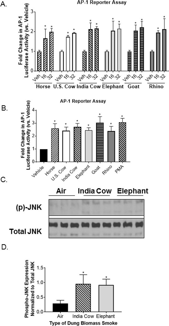
Different types of dung biomass smoke activate JNK-AP-1 in airway epithelial cells.
(A) 16-HBEs were transfected with an AP-1 luciferase reporter and treated with 16 or 32 units/ml of DSE (horse, U.S. cow, India cow, elephant, goat, or rhino) for 24 hours. AP-1 luciferase activity was measured in cell lysates. Data represent mean ± SD (n = 3 replicates per exposure group from an independent experiment), *p<0.05 by two-way ANOVA (compared to vehicle (Veh)-treated cells using Tukey’s multiple comparison post-hoc analysis). (B) AP-1 luciferase activity in 16-HBEs treated with 32 units/ml of DSE. Phorbol myristate acetate (PMA, 50 nM) was included as a positive control. Data represent mean ± SEM (n = 3 – mean of 3 independent experiments), *p<0.05 by one-way ANOVA (compared to Veh-treated cells using Dunnett’s post-hoc analysis). (C) SAECs were exposed to air, Indian cow dung smoke, or elephant dung smoke (30 minutes) and cell lysates were collected 30 minutes post-exposure. A representative Western blot showing 3 technical replicates per exposure group is shown. (C) Densitometry of phospho-JNK and total JNK expression was performed on 3 replicates per exposure group. Data represent mean ± SD (n = 3 replicates per exposure group from an independent experiment), *p<0.05 by one-way ANOVA (compared to air-exposed cells using Tukey’s post-hoc analysis).
