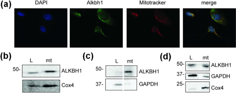
Fig. 2. Subcellular localization of human ALKBH1.

(a) Immunocytochemistry with murine Alkbh1−/− fibroblasts expressing ALKBH1. The DAPI-stained DNA is blue, ALKBH1-bound FITC-conjugated antibody is green, and the mitotracker-localized mitochondria are red. (b) Immunoblot using anti-ALKBH1 and anti-Cox4 antibodies with subcellular fractions from MSU1.1 cells. (c) Immunoblot using anti-ALKBH1 and anti-GAPDH antibodies as in panel b, to confirm the separation of cytoplasmic and mitochondrial proteins (samples are from nonadjacent lanes of the same gel). (d) Subcellular localization of ALKBH1 in HEK293T cells. L, Whole-cell lysate; mt, mitochondria.
