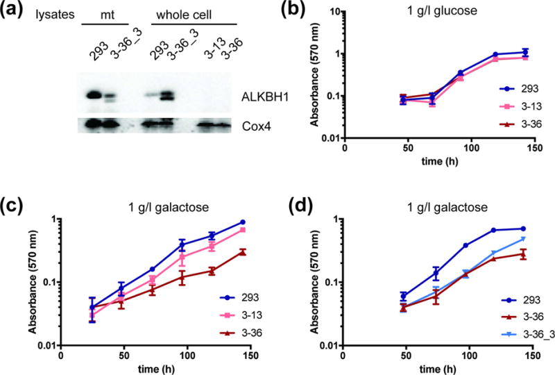
Fig. 3. Representative growth curves of ALKBH1-producing and ALKBH1-deficient HEK293 cells.

(a) ALKBH1 expression in mitochondria and whole-cell lysates of HEK293, ALKBH1-deficient (3–13, 3–36), or Flag-HA-tagged ALKBH1 producing (3–36_3) strains. ALKBH1 expression (endogenous and flag-tagged) was verified in single clones using anti-ALKBH1 antibody, with anti-Cox4 antibody as a loading control. (b) Proliferation of HEK293 cells and two ALKBH1-deficient clones (3–13 and 3–36) in medium containing 1 g/l glucose, measured by MTT staining. (c) Growth of WT and two ALKBH1-deficient strains on 1 g/l galactose, as in panel b. (d) Growth of WT, 3–36 and 3–36_3 cells expressing Flag-HA-ALKBH1 strains on 1 g/l galactose, as in panel b. Data are the mean of four replicates and error bars indicate SD.
