
FIGURE 1. CB3-induced oxidative burst by NOD macrophages is ablated in the absence of NOX-derived superoxide.
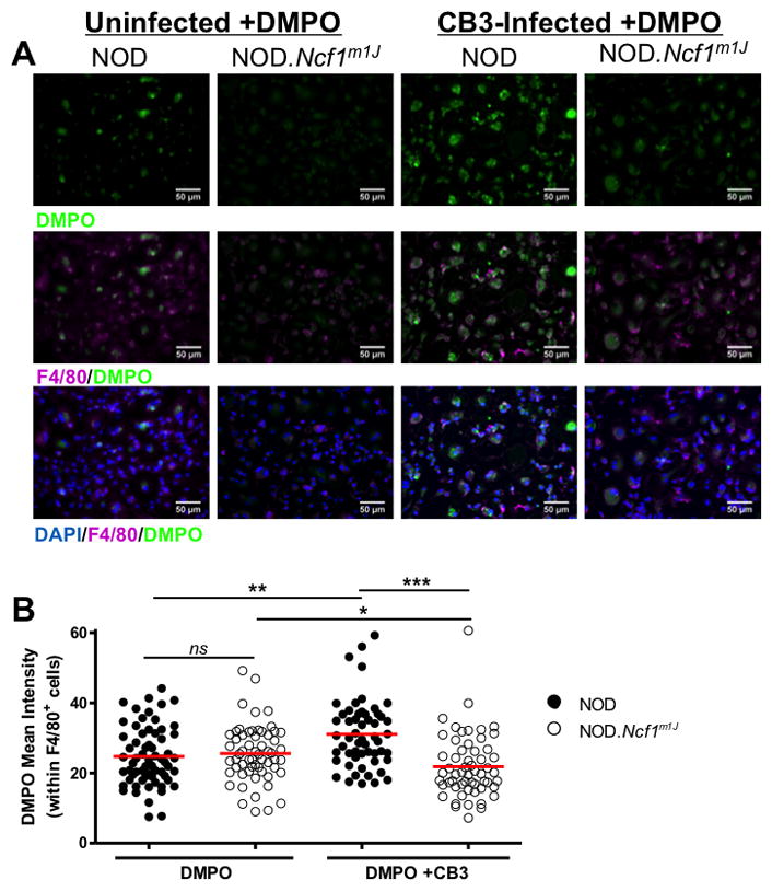
NOD and NOD.Ncf1m1J BM-MΦ were cultured on chamber slides in the presence or absence of DMPO and 10 MOI CB3 infection for 4 hours. Cells were identified by DAPI nuclear stain (blue) and anti-F4/80 macrophage marker staining (purple), and free radical formation of DMPO adducts was detected by anti-DMPO (green) (A). Mean fluorescence intensity of DMPO adducts were quantified using ImageJ analysis software and normalized to F4/80 expression intensity (B). Each dot represents anti-DMPO fluorescence on a single F4/80+ cell with the following total cells quantified: NOD (n=64), NOD.Ncf1m1J (n=59), NOD+CB3 (n=55), NOD.Ncf1m1J+CB3 (n=57). Results are representative of 2 independent experiments. ***p<0.0001; **p<0.01; *p<0.05