
FIGURE 7. Addition of exogenous superoxide restores viral RNA sensor levels and STAT1 activation.
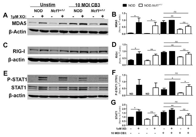
NOD and NOD.Ncf1m1J BM-MΦ were infected with 10 MOI CB3 and co-treated with 1mU/mL XO for 6–24 hours. Whole cell lysates were probed by Western blot for MDA5 (A) and RIG-I (C) at 24 hours, and P-STAT1 (Y701) and STAT1 (E) at 6 hours. Blots were then probed with β-actin as a loading control. Densitometry for MDA5 (B), RIG-I (D), P-STAT1 (Y701) (F) and STAT1 (G) was calculated by normalizing to β-actin and setting expression relative to the NOD CB3+XO treatment group. Western blot images are representative of, and densitometry plots are compiled from, at least 3 independent experiments. **p<0.01; *p<0.05; ns, not significant