Figure 3.
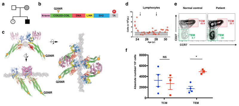
Patient with heterozygous Q206R mutation in STAT5B exhibits accumulation of CD4+ TEM cells in blood.
(a) Pedigree of the affected family: black filled symbol, subject with mutation Q206R; grey filled symbol, subject with mutation Q206R and different clinical phenotype; open symbols, unaffected subjects; (?), unscreened subjects. (b) Schematic representation of the STAT5B protein domain structure. N-term, N-terminus domain (pink); Coiled-coil (CC) domain (green); DNA, DNA-binding domain (red); LINK, linker domain (yellow); SH2, src homology 2 domain (blue). Tyrosine phosphorylation (P, red circle) represented in the transcriptional activation (TA) domain (gray). The Q206R residue is located in the CC domain (orange triangle). (c) Three-dimensional provisional models of STAT5B as a tetramer bound to DNA molecule (gray). Three orientations of the STAT5B tetramer are shown, with Q206R positions indicated as orange circles. (d) Absolute numbers of lymphocytes in the patient’s peripheral blood as a function of age. Arrows indicate times of steroid treatment. (e) Flow cytometry plot of peripheral blood from healthy subject (normal control) and patient, stained for CD27 versus CCR7 (gated on CD4+CD45RA- memory T cells), identifying central memory T cells (TCM: CD27+CCR7+) and effector memory T cells (TEM: CD27-CCR7-). (f) Absolute numbers of TCM and TEM cells per 105 cells of PBMCs from healthy donors (blue) and multiple blood draws from the patient (red). ns = not significant; * p= < 0.05

