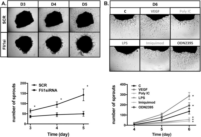
Figure 4. Human dermal ex vivo angiogenesis assay.

A. Dermal tissues obtained from foreskins were treated with SCR or Fli1siRNA for 24h, embedded in matrigel and cultured in EGM-2 medium. B. Dermal tissues were directly embedded in matrigel and cultured with EGM-2 medium. Sprout outgrowth was quantified at day 3,4,5 and 6. Data represent an n=8 well each point with 3 different cultures. Student t-test, *p<0.05
