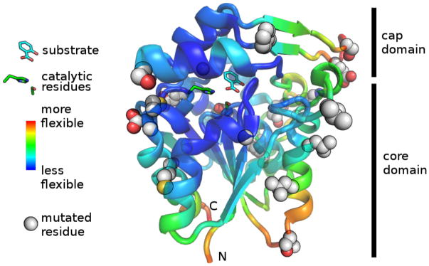
Figure 3.
Location of stabilizing mutations in SABP2. Residues that increased stability when mutated are shown as spheres. The most flexible regions according to B-factors from x-ray crystal structure (yellow/orange/red regions; warmer colors indicate higher B-factor) are the three flexible loops that were targeted for mutagenesis and the C- and N-termini, which were not modified. The product (salicylic acid), as well as catalytic serine (81) and histidine (238), are shown as sticks. The entrance to the active site is from the right in this view. Chain A from pdb:1Y7I30

