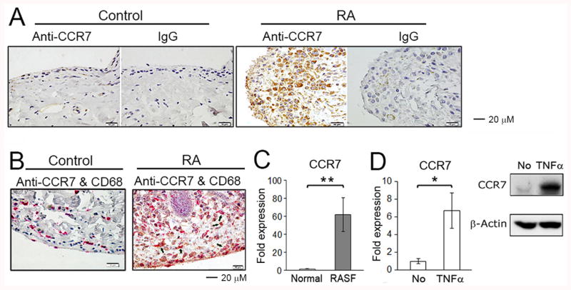
Figure 1. CCR7 is expressed by RA synovial macrophages.

(A) CCR7 expression in arthritis-free control or RA synovial tissue was examined by immunohistochemistry using mouse monoclonal anti-CCR7 and mouse IgG isotype control antibodies. The photos shown are representative of 3 RA and 3 arthritis-free control synovial tissues. (B) The double staining for control and RA synovial tissues employing anti-CCR7 (brown) and anti-CD68 (red) antibodies. The arrows identify cells that are CCR7+CD68+. (C) CCR7 mRNA expression detected by qRT-PCR were examined employing in vitro differentiated macrophages from peripheral blood of 4 healthy donors and RA synovial fluid (SF) macrophages from 9 RA patients. * represents p<0.05 for RA SFs compared to control macrophages differentiated in vitro from normal monocytes. (D) In vitro differentiated human macrophages were incubated for 16 hours with/without TNFα (20 ng/ml). The Expression of CCR7 mRNA was determined by qRT-PCR (left panel) and CCR7 protein expression was detected by Western blot employing an anti-CCR7 antibody (right panel). * represents p<0.05 and ** p < 0.01 between the indicated groups.
