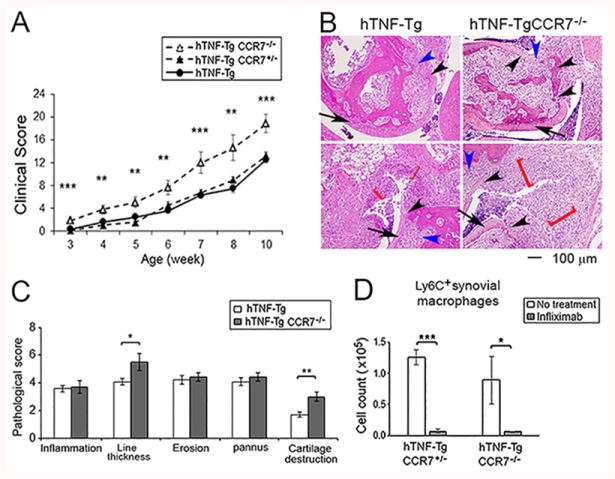
Figure 5. hTNF-Tg CCR7-/- mice develop more severe arthritis but respond similarly to infliximab.

hTNF-Tg mice were crossed with CCR7-/- mice. (A) The development of arthritis in hTNF-Tg CCR7-/-, hTNF-Tg-CCR7+/- or hTNF-Tg (from figure 2A) was evaluated as in Figure 2. The significance was determined by ANOVA among the 3 groups. (B, C). Histological assessment of ankle joints of hTNF-Tg CCR7-/- and hTNF-Tg mice at the same age (n = 5 mice for hTNF-Tg CCR7-/-, and 7 mice for hTNF-Tg mice). The cartilage destruction is identified by the black arrow head and synovial lining thickness by the red bracket, and the inflammation and erosion/pannus were indicated by blue arrow head. The arrows identifies cartilage with normal morphology. (D). Ly6C+ macrophages were determined by flow cytometry and compared between hTNF-Tg-CCR7-/- and hTNF-Tg-CCR7+/- mice. Both groups of mice were treated or untreated with infliximab. The cells were harvested after 3 days following 2 dose of treatment (n = 4-6 mice for each group). * represents p < 0.05, ** p <0.01 and *** p < 0.001 between the indicated groups.
