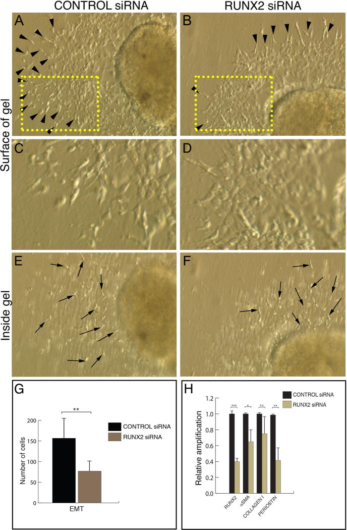
Figure 4. RUNX2-I is required for the activation step of chick heart AVC EMT.
A-F. Stage 14 chicken AVC explants cultured for 24h in the presence of a scrambled siRNA (A, C, E) or a siRNA against RUNX2 (B, D, F). In control explants, multiple activated endothelial cells (arrowheads in A) are present on the gel surface. C represents a higher magnification of the boxed area in A showing the endothelial cell outgrowth. Inside the gel, multiple invasive mesenchymal cells (arrows in E) are observed. RUNX2 siRNA-transfected explants show fewer activated endothelial cells (arrowheads in B) on the gel surface. D represents a higher magnification of the boxed area in B showing a compact endothelial cell outgrowth. Mesenchymal cells (arrows in F) are present in treated explants. G. Quantitation of the number of EMT-derived cells in control and RUNX2 siRNA-transfected explants. There is a significant decrease in the number of activated endothelial cells and mesenchymal cells in treated explants compared to control explants (Average cell number in control explants = 156.71 ± 18.83; average cell number in RUNX2 siRNA-treated explants = 77.00 ± 9.07; ~51% decrease, p < 0.01). n=14 (7, control; 7, experimental). H. qPCR analysis of RUNX2 and mesenchymal cell markers in control and RUNX2 siRNA-transfected explants. There is a significant decrease (~60%) in RUNX2 mRNA after siRNA treatment. RUNX2 downregulation is followed by a decrease in the levels of αSMA (~35% decrease) and PERIOSTIN (POSTN) (~60% decrease). COLLAGEN I is unchanged in treated explants. Error bars = S.E.M.; * p < 0.05; ** p < 0.01; *** p < 0.001; n.s. – non-significant.

