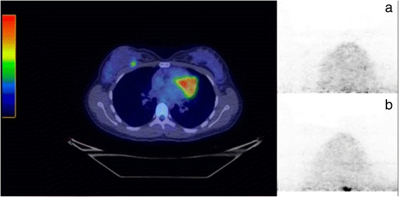
Fig. 8.

PET/CT image of a patient with metastatic lesion from malignant melanoma, located between the ribs and breast. A sub-centimeter focus of increased FDG avidity is seen within the deep soft tissues of the medial right breast. The lesion was not visible on the db-PET image acquired with the standard aperture (a), but can just be visualized at the posterior edge of the images acquired with the medium (200 mm) aperture, with an estimated size of 11.5 mm (b)
