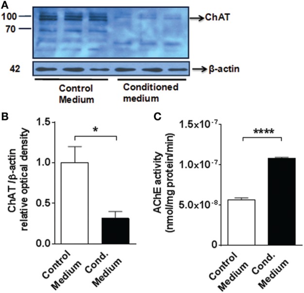
Figure 6.

Medium from LPS-activated microglial cells decreases choline acetyltransferase (ChAT) protein expression and increases acetylcholinesterase (AChE) activity in mouse primary cortical neurons (A) western blot (MW 83 kDa) of ChAT of 7-day-old primary neuronal culture exposed to control or conditioned media (obtained from LPS-activated microglial BV2 cells). See Section “Materials and Methods” for details. (B) Fold change of the western blot determined using Image J to measure band intensities of ChAT normalized to β-actin.*P = 0.0343, Student’s t-test, n = 3 cultures per group, containing neurons obtained from five to six neonate brains each. (C) AChE activity in 7-day-old primary neuronal culture exposed to control or conditioned (Cond.) media ****P < 0.0001, Student’s t-test, n = 3 cultures per group, containing neurons obtained from five to six neonate brains each. The Student’s t-test was performed assuming normality and equal distribution of variance between the groups.
