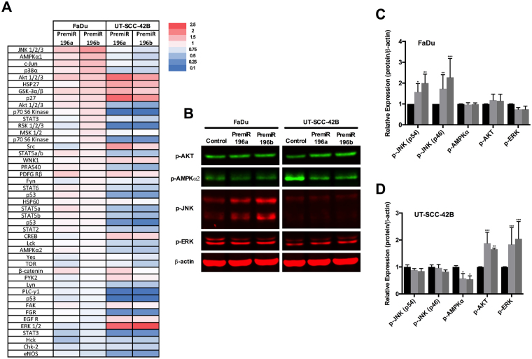
Figure 6.
Analysis of signaling pathways targeted by miR-196a and miR-196b. Total protein lysates from UT-SCC-42B and FaDu cells transfected with either premiR-196a, premiR-196b or non-targeting control were applied to a proteome phospho-kinase array. (A) Relative signal intensity values (to control-transfected cells) are displayed as a heat map. (B) Western blot validation of the array data. (C) and (D) Quantification of IRDye fluorescent signals. *p < 0.05, **p < 0.01 and ***p < 0.001 by Student t-test.

