. 2017 Dec 4;38(2):185–188. doi: 10.3343/alm.2018.38.2.185

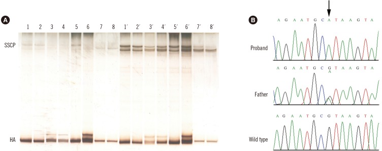
Fig. 1. Heteroduplex, single strand conformational polymorphism, and DNA sequence analyses of the variant region of the ALB gene in the two affected families. (A) Heteroduplex analysis and single strand conformational polymorphism. The DNA encompassing this region of the ALB gene of four members of the Turkish family, two members of the Algerian family, and two controls was amplified with the PCR primers A19B and A20C [4], and the fragments were electrophoresed onto a non-denaturing polyacrylamide gel. Turkish family: lane 1, patient 2; lane 2, patient 1; lane 3, mother; lane 4, father. Algerian family: lane 5, patient, and lane 6, father. Lanes 7–8, controls. The same samples were denatured and cooled before loading. Turkish family: lane 1´, patient 2; lane 2´, patient 1; lane 3´, mother; lane 4´, father. Algerian family: lane 5´, patient and lane 6´, father. Lanes 7´–8´, controls. (B) Genomic DNA sequence electropherograms of the variant region of the ALB gene in the Algerian family. The arrow indicates the G>A transition at position c.1289+1, the first base of intron 10. The patient is homozygous for the mutation, while the father is heterozygous for the wild-type and mutated alleles, as evidenced by the presence of two superimposed peaks.

Fig. 1