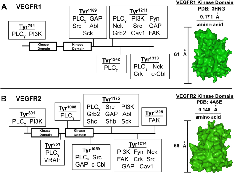
Fig. 1.
VEGFR–adapter interaction schematics. This schematic depicts the known biology of VEGFR1, VEGFR2, and their related adapters. Adapters bind specific tyrosine (Tyr) sites on a VEGFR1 and b VEGFR2 (SI Table S4). VEGFR1 and VEGFR2 kinase domain crystal structures were used to measure the distance between individual VEGFR amino acids. This measurement, along with adapter size measurements (SI Table S5), were used to map the adapters and Tyr sites that allow multiple adapters to bind a VEGFR simultaneously, as described in the Supplementary Information

