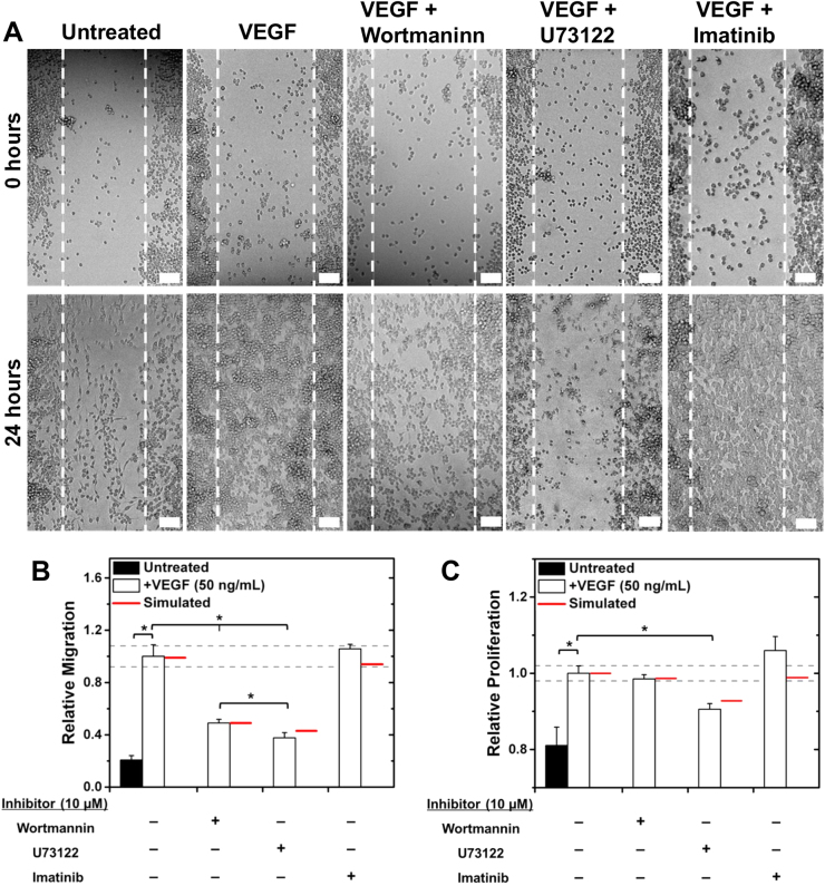
Fig. 5.
PLCγ and PI3K regulate VEGFR1-induced cell responses in vitro. a RAW migration was measured in wound-healing assays at 0 and 24 h post scratch. Scale bars represent 50 µm. b Analyzed wound-healing assays show that inhibiting PLCγ or PI3K significantly decreases VEGF-induced RAW migration. c PLCγ inhibition significantly decreases VEGF-induced RAW proliferation, measured with MTT assays. Treatments for all experiments were: 50 ng/mL VEGF-A164, 10 µM Wortmannin (PI3K inhibitor), 10 µM U73122 (PLCγ inhibitor), and 10 µM Imatinib Mesylate (Abl inhibitor). All experiments were performed in triplicate, and data are represented as mean ± SEM. Experimental significance is given at p < 0.05. (b, c) The predicted maximum reduction in cell response is given for each inhibitor treatment (red line) using the physiologically specific VEGFR tyrosine ite model. Dashed gray lines outline the range corresponding to 10% variation in cell migration given VEGF treatment alone; inhibitor treatments are predicted to be significant by the model if the predicted cell migration lies outside this range. Model predictions include cell response contributions from both VEGFR1 and VEGFR2 for validation purposes

