
Figure 4.
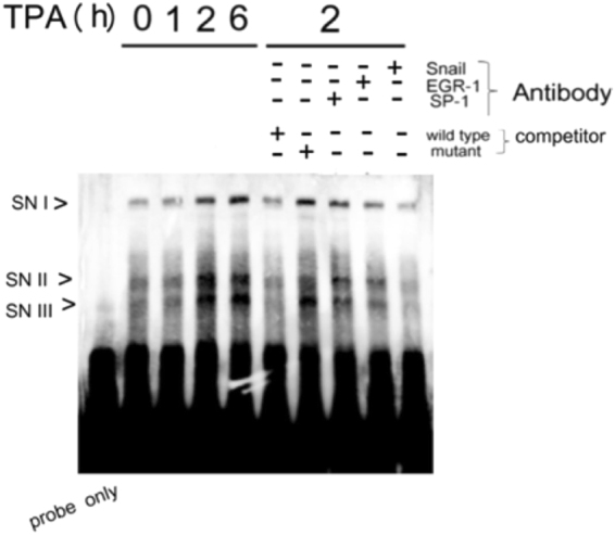
EMSA for in vitro binding of Snail to the proposed Snail binding region on MMP9 promoter. Nuclear extracts obtained from HepG2 were treated with 50 nM TPA for the times indicated, followed by EMSA using MMP9-proSN (lanes 2-5). Unlabed wild-type or mutant competitors in 200X amount were included in the EMSA for HepG2 treated with TPA for 2 h (lanes 6 and 7). Lane 1 is the sample of probe only. For antibody blocking analysis, each of the indicated antibody was preincubated with the nuclear extract from HepG2 treated with TPA for 2 hr followed by EMSA reaction (lanes 8–10). Schematic map of the EMSA probes (MMP9-porSN), and the competitor (MMP9-porSN) located around Snail binding motif upstream of the EGR-1/SP1 overlapping region on MMP9 promoter is demonstrated in supplemental Fig. 2C.