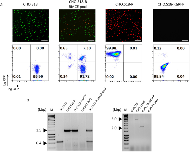
Figure 2.
Drug selection-free RMCE. LPV-encoded GFP reporter genes in CHO cells were replaced by the DVRMCE sequences. (a) Representative fluorescence microscopy images and flow cytometry plots of, from left to right, the CHO.S18 master clone, the unselected RMCE pool after the RMCE transfection, an isolated RFP+ clone (CHO.S18-R) and the same clone after Cre-mediated RFP gene deletion (CHO.S18-RΔRFP). Scale bar = 100 µm. (b) Verification of the expected RMCE (left) and deletion events (right) by PCR analysis of genomic DNA. The nomenclature of the cell lines and pools is according to Fig. 2a, above. Shown on the left is a multiplex PCR with 3 primers where the lower band of 537 bp is indicative of the intact LPV, whereas the upper band of 1,329 bp is indicative of the RMCE reaction. Shown on the right is a PCR analysis of integrated DVRMCE, before (upper band of 4,571 bp) and after (lower band of 2,157 bp) Cre/loxP deletion. For details of the PCR patterns refer to suppl. Fig. S9. (M: Hyperladder 1 kb). The uncropped gel pictures are shown in S11a.

