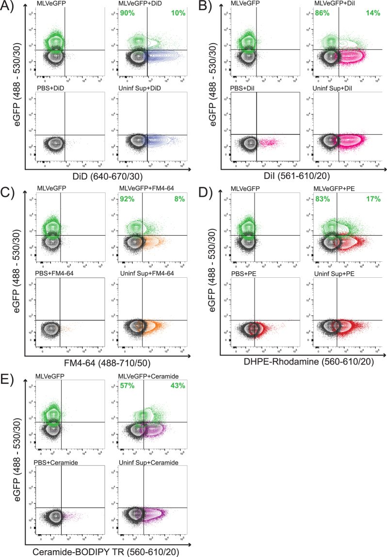
Figure 4.
Direct staining of virus. MLVeGFP viruses from chronically infected NIH 3T3 cell supernatants were labeled with (A) DiD, (B) DiI, (C) FM4–64, (D) Rhodamine-1,2-DihexadecanoylPhosphatidylethanolamine (DHPE-Rhodamine), and (E) Ceramide-BODIPY TR. Samples were then analyzed by NFC without purification. eGFP+ particles are depicted in green, background noise or non-fluorescent particles in black, dye-labeled particles in color (other than green). The distribution of total dye-labeled and unlabeled eGFP+ particles is indicated in green. Included in each panel: unstained virus (MLVeGFP), stained virus (MLVeGFP+ dye), dye alone (PBS + dye), and uninfected supernatant with dye that serves as an EV-only sample (Uninf Sup + dye).

