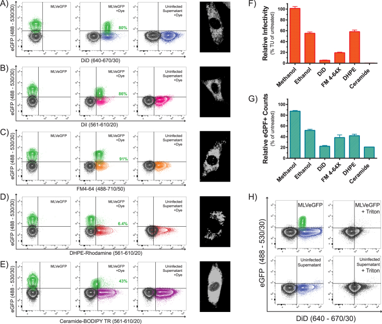
Figure 5.
Indirect staining of virus. Supernatants of chronically infected and uninfected cells labeled with (A) DiD, (B) DiI, (C) FM4-64, (D) DHPE-Rhodamine, and (E) Ceramide-BODIPY TR were analyzed by NFC. (F) Relative virus infectivity (transducing units (TU) per mL) and (G) counts of eGFP+ particles released from the supernatants of infected cells labeled with the different dyes. Infectivity and counts are presented as the percentage of total number of eGFP+ particles released from unstained virus-infected cells. Data depicts the mean with S.D. from three independent experiments. (H) Supernatants from DiD-stained infected and uninfected cells were treated with 0.05% Triton X-100.

