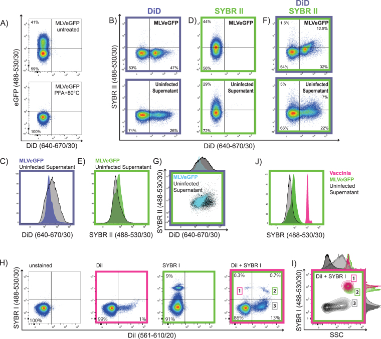
Figure 6.
Discrimination of enveloped viruses from EVs by a combination of membrane and nucleic acid dyes. (A) MLVeGFP was treated with 2% PFA at 80 °C to fix the virus and permeabilise the envelope and capsid, rendering the content of the particles amenable to staining by nucleic acid dyes. MLVeGFP infected and uninfected cells were stained with DiD, and then particles in the supernatant were analysed directly (B), or further stained with SYBR II following fixation and heat treatment (F). (D) Supernatants from infected and uninfected cells were stained with SYBR II only and analysed. Comparison of fluorescent intensities between supernatants from infected (coloured histograms) and uninfected cells (black histograms): (C) DiD stained cells, (E) SYBR II stained supernatants, (G) histogram and dotplot overlay of dual stained DiD/SYBR II nanoparticles from infected and uninfected cells. Nanoparticles from uninfected cells are depicted in black, and from infected cells in blue. (H) Direct labeling of VV with DiI or SYBRI alone, or in combination (fourth panel). (I) Relative sizes of nanoparticle populations 1 to 3, displayed as a comparison of SYBRI vs SSC. (J) Overlay of SYBR fluorescence intensity from stained MLVeGFP, VV and EV-containing cell supernatant.

