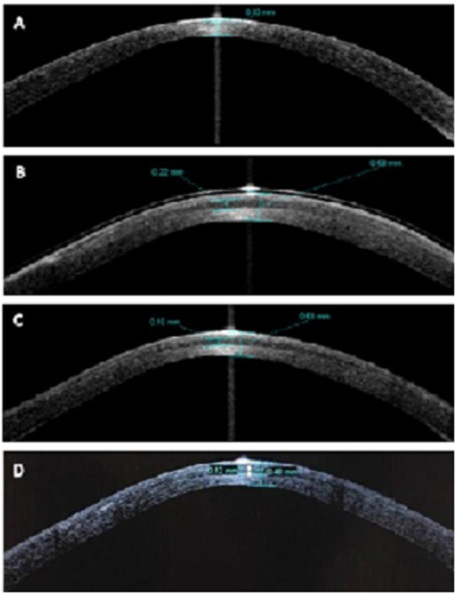
Figure 3.

Optical coherence tomography images of the cornea in post laser-assisted in situ keratomileusis (LASIK) ectasia patient (patient 3). (A) Preoperatively, the residual corneal thickness was only 330 μm. (B) The total corneal thickness was 580 μm, while corneal lenticule thickness was 220 μm on the first day after surgery. The interface between the corneal lenticule and the stroma bed was clear. (C) The total corneal thickness was 530 μm, while corneal lenticule thickness was 160 μm in 2 weeks after the surgery. (D) The total corneal thickness was 480 μm, while corneal lenticule thickness was 120 μm at 12 months after the surgery. The interface between the corneal lenticule and the stroma bed became ambiguous.
