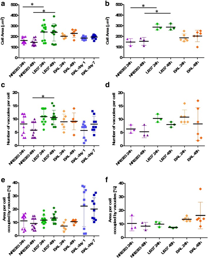
Fig. 2.
Median morphology parameters of untreated and amiodarone-treated rat and human macrophage cell models. Median cell area, vacuole number per cell and percentage of cell area occupied by vacuoles for untreated cells (a, c, e) and cells exposed to 10 μM amiodarone (b, d, f). NR8383 rat macrophage cells (purple triangle), U937 human monocyte-derived macrophage cells (green circles), primary rat macrophages derived from BAL in in vitro culture for 24 h or 48 h (orange diamonds) and primary rat macrophages derived from BAL tested immediately after harvesting from naïve rats after 1 and 7 day timepoints (blue squares). Each data point represents the median value of n = 6 wells per plate. * indicates p < 0.05.

