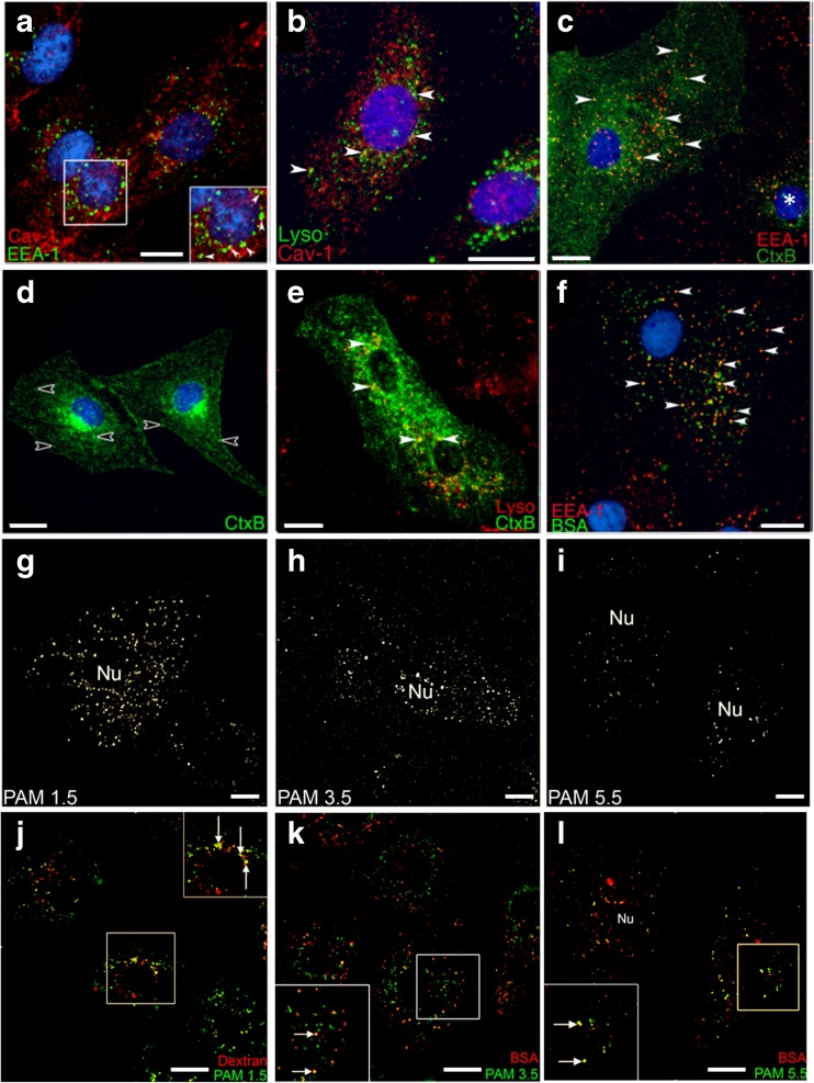
Fig. 2.
Distribution and functionality of endocytic compartments in ATI-like lung epithelia. Co-localisation of Caveolin-1 positive vesicles with (2a) EEA-1 immunopositive endosomes and (2b) lysosomes labelled by a 2 h/3 h pulse-chase internalization of dextran. CtxB internalized into ATI-like cells localizes to (2c) early endosomes after 10 min, (2d) a peri-nuclear compartment after 45 min. (2e) only a minor fraction of CtxB puncta localized to lysosomes following a 2 h/3 h pulse-chase co-internalisation of CtxB and 10 kDa dextran. Empty arrowheads in 2D highlight non-perinuclear puncta present within upstream compartments. (2f) TMR-labelled BSA accumulated in EEA-1 positive endosomes after a 10 min incubation. (2g–I) Primary ATI-like cells show a decrease in the density of PAMAM-OG staining density pattern that accompanies an increase in dendrimer generation from 1.5 to 5.5 after 30 min incubation. (j) a fraction of PAMAM 1.5 co-localised with dextran labelled lysosomes after a 2 h/3 h pulse-chase co-internalisation. Both PAMAM 3.5 (k) and PAMAM 5.5 (l) co-localise with BSA after 30 mins co-internalisation. Arrows in zoomed section highlight co-localised puncta. Scale bar: 5 μm.

