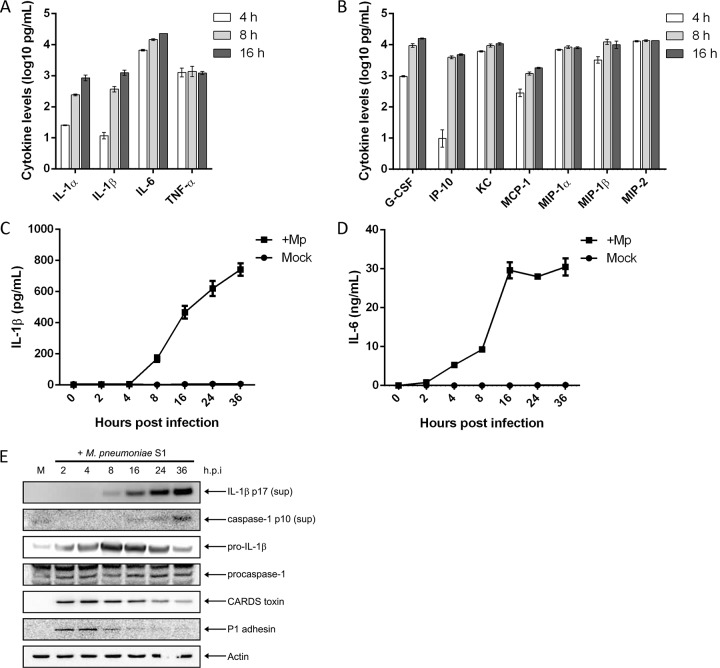
FIG 1.
Primary macrophages infected with M. pneumoniae produce proinflammatory and chemotactic cytokines. WT BMDMs were infected with M. pneumoniae at an MOI of 100 mycoplasmas per cell. At 4, 8, and 16 h postinfection, cell culture supernatants were collected and used in multiplex cytokine bead assays. Values for proinflammatory cytokines (A) and other cytokines and chemokines (B) were normalized by subtracting values of the uninfected control from those of the infected animals and are represented in log scale. Time course kinetics for IL-1β and IL-6 secretion are shown in panels C and D, respectively. (E) Immunoblot analysis of IL-1β p17 and caspase-1 p10 in cell culture supernatants (sup) and pro-IL-1β, procaspase-1, CARDS toxin, and P1 adhesin in cell lysates of WT BMDMs infected with M. pneumoniae. The mock-infected control is designated M. β-Actin served as the loading control. Data in panels A and B are representative of two independent experiments with similar results; data in panels C and D are representative of two independent experiments with similar results. h.p.i., hours postinfection; Mp, M. pneumoniae. All cytokine and chemokine levels are listed in Table S1 in the supplemental material.

