FIG 3.
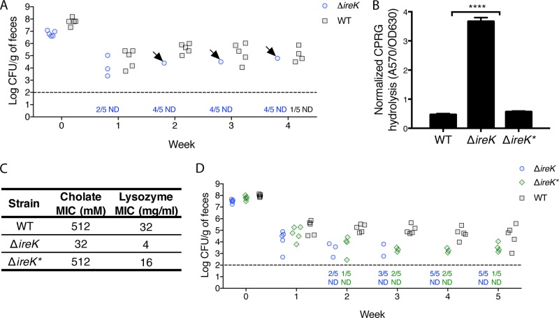
The intestinal environment can select for suppressor mutants that recover envelope integrity, antimicrobial resistance, and the ability to colonize. (A) Groups of mice (5 per group) were colonized with either WT E. faecalis (OG1RF) or the ΔireK mutant (CK119). Colonization levels were determined by enumerating the enterococci in feces by culture on rifampin-supplemented BHI agar. Arrows, mice harboring ΔireK* clones; dashed line, the limit of detection. Symbols for mice with undetectable colonization levels were omitted, and instead, the number of mice for which colonization was not detected (ND)/total number of mice tested is shown underneath the dashed line. (B) CPRG hydrolysis was measured for the ΔireK* suppressor mutant, the WT, and the ΔireK mutant. Reported measurements represent averages from three independent cultures, and error bars represent standard deviations. Statistical significance was evaluated by t test. ****, P < 0.0001 versus WT. (C) The cholate and lysozyme resistance of the strains listed in panel B was determined. The reported MICs represent the median values from three independent biological replicates. (D) Groups of mice (5 per group) were colonized with either WT E. faecalis (OG1RF), the ΔireK mutant (CK119), or the ΔireK* suppressor mutant. Intestinal colonization was assessed as described in the legend to panel A.

