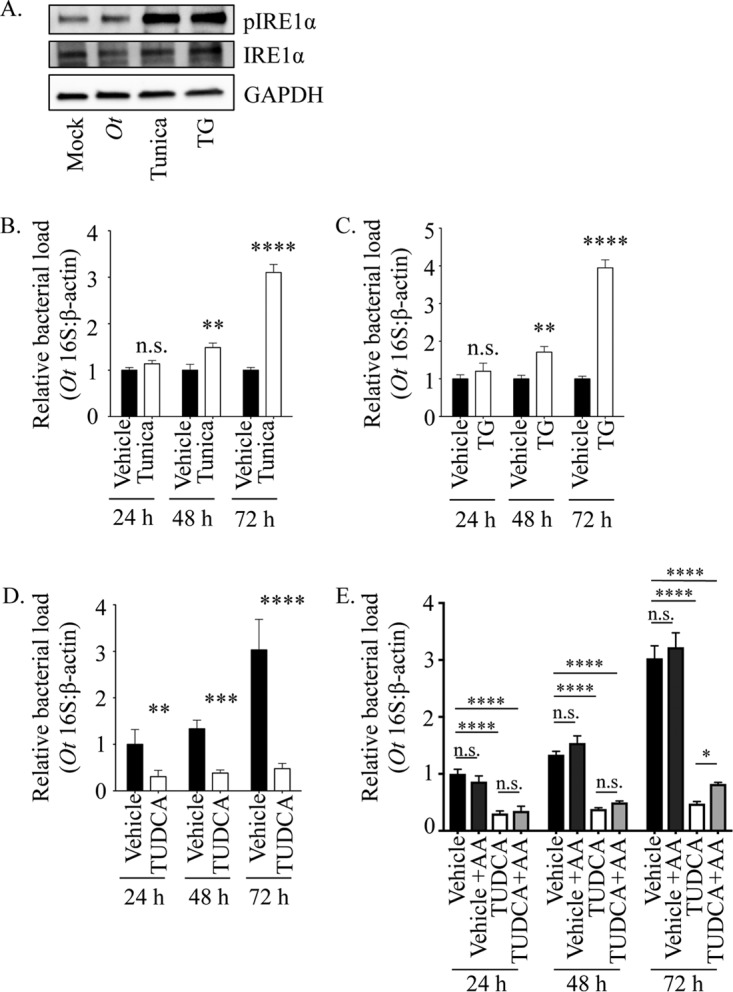
FIG 2.

O. tsutsugamushi benefits from ER stress in an amino acid-dependent manner. (A) Whole-cell lysates of HeLa cells that were mock infected, O. tsutsugamushi infected (Ot), or treated with tunicamycin (Tunica) or thapsigargin (TG) were analyzed by Western blotting and screened with antibodies specific for phosphorylated IRE1α, IRE1α, or GADPH. (B to D) HeLa cells were treated with tunicamycin (B), TG (C), TUDCA (D), or vehicle for 1 h prior to and throughout infection with O. tsutsugamushi. Total DNA isolated at 24, 48, and 72 h was analyzed by qPCR. Relative levels of the O. tsutsugamushi 16S rRNA gene were normalized to the relative levels of β-actin by using the 2−ΔΔCT method. Resulting relative levels per treatment were normalized to those of the respective vehicle controls at each time point. (E) The TUDCA experiment was performed as described for panel D, except that an additional sample was included in which vehicle- or TUDCA-treated O. tsutsugamushi-infected HeLa cells were grown in medium containing amino acids (+AA). Statistically significant values are indicated: *, P < 0.05; **, P < 0.01; ***, P < 0.001; ****, P < 0.0001; n.s., not significant. Results are representative of at least three separate experiments performed in triplicate.
