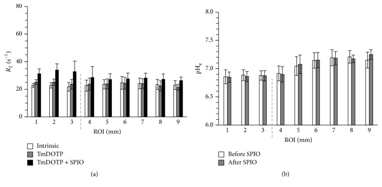
Figure 3.
Region of interest (ROI) analysis for the average relaxation rate (R2) and extracellular pH (pHe) before and after infusion of SPIO-NPs for all RG2 tumor-bearing rats that underwent coinfusion of TmDOTP5− and probenecid (n = 5). The ROI analysis is based on concentric 1 mm circular rings drawn from the center of mass of the tumor. (a) Average R2 values in different ROIs for intrinsic contrast, after infusion of TmDOTP5−, and after infusion of TmDOTP5− with SPIO-NPs. The average R2 enhancement was highest inside the tumor (ROIs 1–3) compared to tumor edge (ROI 4) and nontumor regions (ROIs 5–9). The dashed line represents the tumor edge. Small R2 enhancement was observed after infusion of TmDOTP5−. However, much higher R2 enhancement was observed upon infusion of SPIO-NPs. See Table 1 for details. (b) The average pHe values in different ROIs before and after infusion of SPIO-NPs. The pHe values measured before and after infusion of SPIO-NPs were similar, both inside and outside the tumor. The pHe was lowest in the tumor and highest in the healthy/nontumor tissue farthest from the tumor. Low pHe was also measured on the tumor margin. See Table 2 for details.

