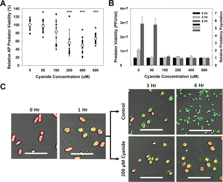
FIG 4 .
(A) Cyanide is mildly, yet significantly, toxic to attack-phase B. bacteriovorus HD100. B. bacteriovorus cultures were exposed to different micromolar concentrations of cyanide for 24 h, after which the viable populations were determined. A total of 12 independent cultures were tested. Each symbol represents the value for one culture, and the average result for a particular cyanide concentration is indicated by a light gray circle. Values were compared to the value for the no-cyanide control samples. Values that are significantly different (P < 0.001) are indicated (***). This experiment was repeated 12 times. (B) Cyanide prevents release of the predatory cells from the bdelloplast. This graph shows the numbers of viable predators, which include both free-swimming predators and those within the bdelloplast, seen during and after a single predation event. The relative number illustrates the titer burst seen when lysis of the bdelloplast occurs. This experiment was repeated six times. (C) Confocal microscopic images showing the impact of cyanide on the development of the predatory cells within the bdelloplasts. E. coli MG1655 prey express the red fluorescent protein (red), while the predator is expressing the Venus yellow fluorescent protein (green). In the presence of 200 µM cyanide, elongation and development of the intraperiplasmic predatory cells, and the resulting lysis of the bdelloplast, are halted. This agrees with the results shown in panel B. The 0-h image was taken just prior to introducing the predatory strain. Bars = 10 µm.

