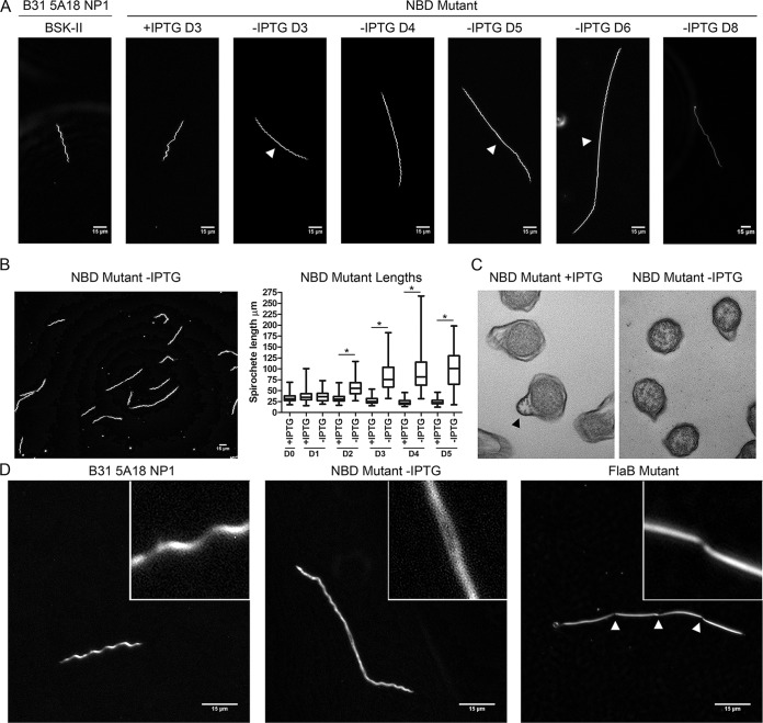
FIG 5 .
Peptide starvation induces dramatic morphological changes during in vitro cultivation. (A) Results of dark-field microscopy (magnification, ×400) showing the wild-type strain (B31 5A18 NP1), the NBD mutant with (+) IPTG and without (-) IPTG. D3 of the wild-type strain and NBD mutant with IPTG is representative of all time points. Arrowheads designate central flattening in mid-cell regions of the NBD mutant without IPTG. (B) Representative image (magnification, ×400) of the conditional NBD mutant without IPTG on D4 post-depletion along with whisker plots of spirochete lengths measured using the Metamorph integrated morphometry analysis tool. Asterisks (*) denote P values of <0.0001 for pairwise comparisons at each time point determined using a two-tailed t test. (C) Transmission electron microscopy cross sections demonstrating diminished flagellation in organisms incubated without IPTG. Flagellar bundles are denoted with an arrowhead. (D) Dark-field microscopy (magnification, ×1,000) of the wild-type strain, a conditional NBD mutant without IPTG (D5), and a ΔflaB mutant. Septal invaginations in the ΔflaB mutant are denoted with arrowheads.

