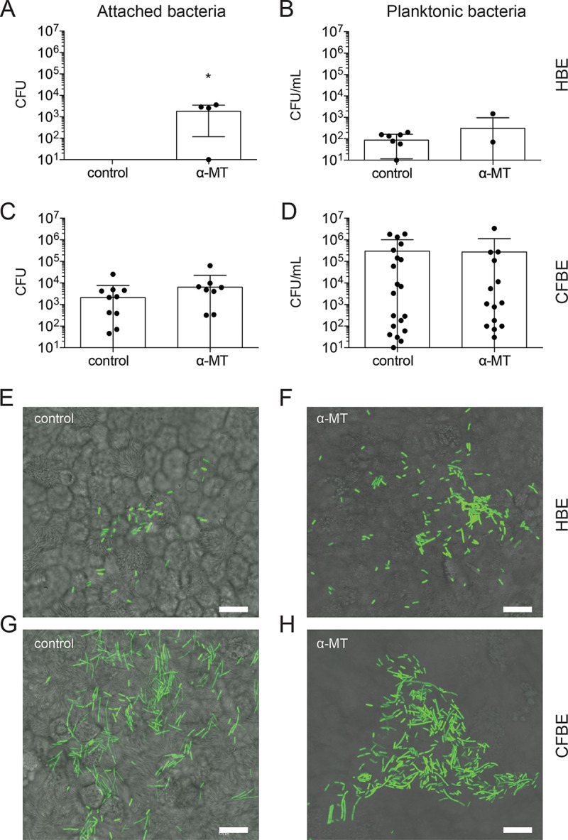
FIG 5 .

Inhibition of SLC6A14 results in increased bacterial attachment to HBE monolayers. Quantification of bacterial CFU attached to the epithelial cell monolayer or collected from the infection medium (planktonic). (A) Treatment of HBE cell monolayers with α-MT results in a significant increase in the amounts of attached P. aeruginosa recovered after 8 h of infection (P < 0.05). (B) Viable bacterial counts in infection medium show that treatment with α-MT does not have a significant effect on planktonic growth of P. aeruginosa in HBE cell cocultures. (C and D) Treatment with α-MT did not influence attached or planktonic bacterial cell counts in CFBE cocultures. Individual data points are shown for biological replicates with CFU counts above the lower limit of detection ± the standard deviation. Numbers of patient samples with CFU counts below the detection level: A, 9 and 1; B, 2 and 3; C, 11 and 7; D, 1 and 1 (control and treated, respectively). Asterisks indicate a P value of <0.05 (two-tailed unpaired t test assuming equal standard deviation [B and D], not assuming normal distribution [A, Mann-Whitney test], or not assuming equal standard deviation [C, Welch’s correction]). (E to H) Epithelial cell monolayers were imaged by confocal microscopy at 8 h postinfection. Higher numbers of GFP-expressing P. aeruginosa bacteria can be seen on the surface of HBE cells treated with α-MT than on that of untreated control HBE cells. CFBE cell monolayers show a greater bacterial burden than HBE cells; however, α-MT did not have an appreciable effect on the quantity of bacteria visualized on the surface of CFBE cell monolayers. Scale bars, 10 μm.
