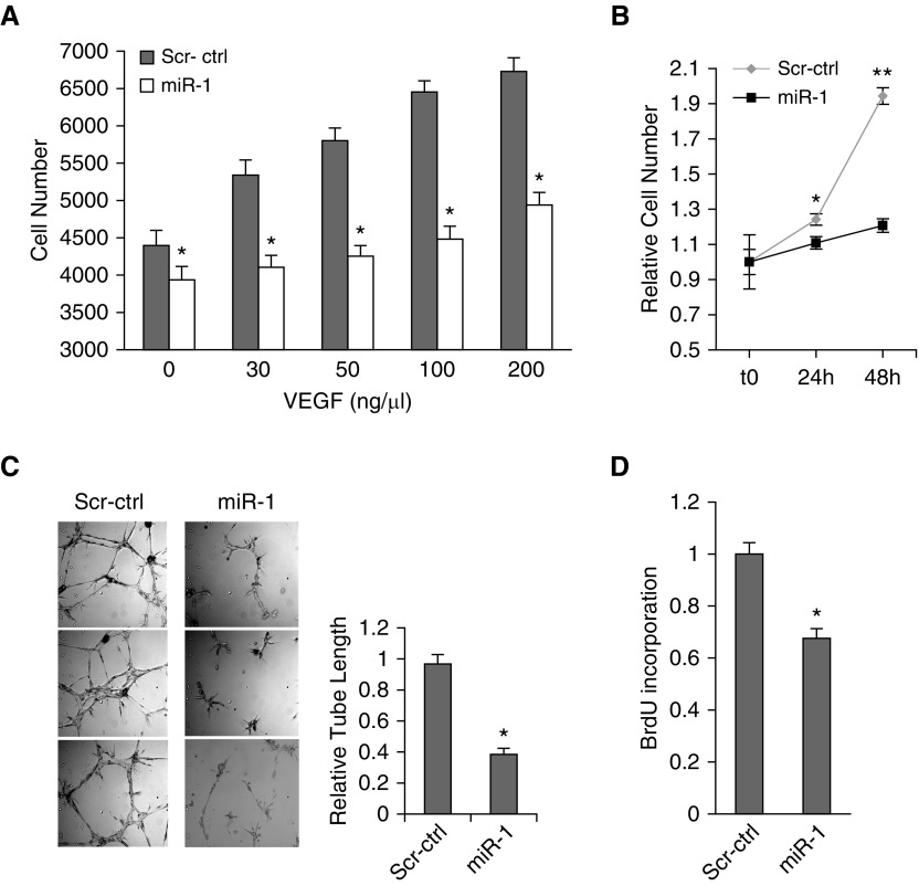
Figure 5.
The effect of microRNA-1 on mouse lung endothelial cells (MLEC). (A–D) MLECs were transfected with double stranded miR-1 mimic (miR-1) or scrambled control RNA (Scr-ctrl) and stimulated with recombinant human vascular endothelial growth factor (VEGF)-A. (A) MLEC growth at 24 hours in response to various concentrations of VEGF was measured using WST-1 kit (Roche) (n = 6 from two experiments; *P < 0.05). (B) MLEC growth in response to 50 ng/ml of VEGF at the indicated time points was measured as in A (x-axis shows time) (n = 24 from four experiments; *P < 0.01, **P < 0.000001). (C) Endothelial sprouting (capillary tube formation in matrigel). (Left) Representative images. (Right) Quantitation of the relative tube length (n = 9 from three experiments; *P < 0.005). (D) Bromodeoxyuridine incorporation in response to VEGF was measured as an index of de novo DNA synthesis by ELISA (Roche) and values were normalized to the control reaction (n ≥ 24 from six experiments; *P = 1.4 × 10⁻⁶). Error bars represent SEM. BrdU = bromodeoxyuridine.

