Figure 6.
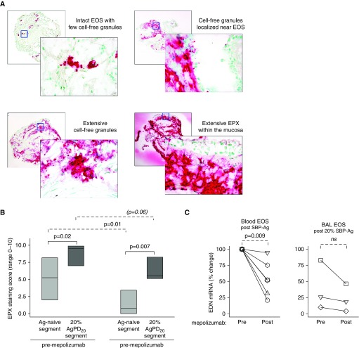
Eosinophil release of eosinophil peroxidase (EPX) and EPX-containing granules is reduced, but still present, in bronchial mucosa after mepolizumab administration. (A) Representative photomicrographs, taken using a ×10 objective (rear photographs) and a ×40 objective (forward photographs), demonstrating EPX staining patterns. (B) After mepolizumab administration, the EPX staining score (medians with first and third quartiles) was significantly less for the antigen-naive site and tended to decrease for the site that had been challenged with 20% of the participant’s AgPD20 (allergen provocation dose resulting in a 20% reduction in FEV1) (n = 8). (C) EDN mRNA fold change normalized to each individual’s premepolizumab circulating eosinophils. Symbols represent values for individual subjects (n = 6 for blood eosinophils; n = 3 for BAL eosinophils). Pre– versus post–SBP-Ag P values are indicated above the solid lines; pre- versus postmepolizumab P values are indicated above the dashed lines. Ag = antigen; BAL = bronchoalveolar lavage; EDN = eosinophil-derived neurotoxin; EOS = eosinophils; ns = not significant; SBP-Ag = segmental bronchoprovocation with allergen.

